Baby Foot Sticking Out of Uterus True or False
Baby's Feet Exterior Mom'south Uterus: Astonishing Image Shows Rare Rupture
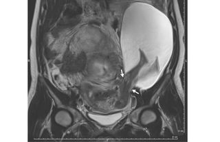
Merely looking at this image might give the impression that this woman'due south baby literally kicked its feet right out of her uterus. Simply moms-to-be with kicky babies tin can residual easy — the MRI image showcases an extremely rare condition that was not caused by a babe'southward boot.
The 33-yr-old adult female had developed a 1 inch (2.5 centimeters) tear in the wall of her uterus, and through the tear, part of the amniotic sac measuring 7.five by 4.7 by 3.5 inches (nineteen by 12 by 9 cm) popped out, co-ordinate to a cursory report of her case. The amniotic sac is the fluid-filled membrane found in the uterus that contains the growing and developing fetus.
But the woman had no symptoms that any of this was going on. She didn't learn of her condition until she came in for a routine ultrasound when she was 22-weeks pregnant, according to the report, published today (Dec. 21) in The New England Journal of Medicine.
Dr. Pierre-Emmanuel Bouet, an OB/GYN at the Angers Academy Infirmary in France and the lead writer of the report, said he had never seen a case similar this earlier. [Here's a Behemothic Listing of the Strangest Medical Cases We've Covered]
Indeed, the condition is "extremely rare," Bouet told Live Science. There have simply been 26 cases reported in the literature, he added.
This was the woman'south sixth pregnancy, the doctors wrote in the written report. In all of her five previous pregnancies, the adult female delivered the babies via Caesarean department (C-section), they wrote.
In fact, it was the woman'south five previous C-sections that increased her risk for a uterine tear, Bouet said. It seems that her C-sections had weakened the wall of the uterus, he said. The tear didn't occur at the exact location of the earlier C-sections, simply close by, he added.
The area of the uterus that had scarred after the C-sections was strong, simply the regions around this scar were fragile, Bouet said. The forces and pressures on the uterus that occur during pregnancy ultimately led to the tear, he said.
Upon discovering the woman'south uterine tear and protruding amniotic sac, the doctors informed the adult female and her married man of the potential risks, which included additional uterine tearing, preterm nascence and a serious pregnancy complication called placenta accreta, in which the placenta doesn't detach from the uterine wall after birth.
It was too possible for the amniotic sac to rupture, Bouet said. If this occurred, the doctors would brand certain that the fetus nonetheless had a heartbeat, and if so, would perform an emergency C-section, Bouet said. The doctors would also have to consider the historic period of the fetus: if it was too early in the pregnancy, the odds of survival would exist lower, he said.
The adult female and her husband decided to continue the pregnancy with shut monitoring, co-ordinate to the study. Bouet said that the woman was non put on bed rest during this time, and that she could do some moderate walking.
By 30 weeks, the tear in the woman's uterus grown past 2 inches (5 cm) and the portion of the amniotic sac outside the uterus had grown in size, the doctors wrote in the report. At that point, not merely did this role of the amniotic sac incorporate the fetus's legs, just also the abdomen, they wrote.
The doctors and the woman decided to evangelize the baby via C-section. The baby boy was healthy, and weighed in at 3 lbs. (1.385 kilograms), according to the report. Later on delivery, the doctors repaired the woman'southward uterus, and she returned dwelling house from the infirmary subsequently five days.
The doctors concluding checked in with mother and baby half dozen months after he was built-in, and noted that they were doing well.
Originally published on Alive Science.

Sara is a staff writer for Live Science, covering wellness. She grew upwardly outside of Philadelphia and studied biological science at Hamilton Higher in upstate New York. When she's not writing, she tin can be institute at the library, checking out a big stack of books.
Source: https://www.livescience.com/57292-uterine-rupture-amniotic-sac.html
Post a Comment for "Baby Foot Sticking Out of Uterus True or False"